Module list of functions Versana Premier
Whizz Image Tuning
With a single button press, Whizz continuously and automatically optimizes image quality as you move between different organs or structures—no repeated adjustments required.
Scan Assistant
Provides predefined exam protocols that guide you through each imaging step—reducing keystrokes, stress, and fatigue while maximizing consistency.
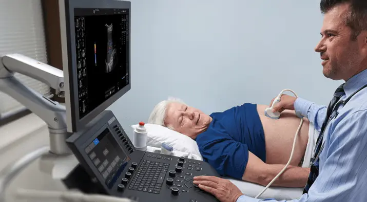
AutoBladder in Whizz
Combines optimized ultrasound imaging with automated bladder measurements for quick, accurate evaluations
Breast Care
Perform breast examinations with a systematic approach that ensures coverage of all segments, conducting evaluations step by step. For serial exams, compare images to reassess the quadrants previously marked as risks.
Auto EF
Automatically tracks myocardial tissue strain and calculates left ventricular ejection fraction (LVEF) from apical views, simplifying cardiac function assessment.
Needle Recognition
Improves visibility of needle tips for precise biopsies, enhancing patient safety and clinician confidence.
Voice Annotations
Connect a microphone, tap an icon, and capture hands-free voice notes overlaid on images—reviewable later for comprehensive exam documentation.
Productivity of the breast
Systematic breast scans with integrated BI-RADS comments and auto contouring tools help you document findings and measure structures easily.
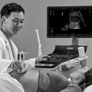
SonoBiometry
Suggests caliper placements for standardized fetal measurements (BPD, HC, AC, FL, HL) in OB scans, boosting exam efficiency.
Insite Technology
Enables remote service and diagnostics via GE’s advanced monitoring platform—resolving many issues instantly without onsite visits.
Stress echocardiography
An editor template for the stress testing of the myocardium. To evaluate the function of the segment heart by the score and comparison of the effort of the myocardium. The stress echocardiogram allows the scoring of wall motion and the automatic labeling of the level of effort of the measurements.
Productivity of the thyroid
To evaluate the thyroid as it performs scanning; describe the characteristics of the structure with comments that are directly incorporated in the report.
VOCAL
Analysis of virtual organ computer-aided. 3D tool that allows the calculation of the volume of areas of eccentrically or complex anatomical structures.
Tissue Doppler & TVI
Acquire data in the background during a scan 2D normal.
B-Flow and B-Flow Color
B-Flow is intended to provide a more intuitive
the hemodynamic not quantitative in vascular structures.
Ultrasound contrast* enhanced (CEUS)
Technique for the detection of vascularization that uses the properties of reflection of the sound of the contrast agent injected.
Image acquisition speed tissue (TVI, Tissue Velocity Imaging)
Estimated and encodes the speed of the tissues by color.
3D/4D
To visualize the three planes of the image in a volume of exploration in real-time; this is especially useful in tests obstetric. The representation and visualization of 3D images provide a rapid reconstruction of scans and anatomical. The 4D technology provides a continuous acquisition and high-volume 3D images. The 4D technology adds a dimension of movement to a 3D image.
Effective follow-up examination
In the follow-up examinations, the tracking tool automatically identifies the same ultrasonic probe, presets, depth, gain, and frequency were used in the previous exploration.
The current picture of ultrasound in real-time is shown next to the previous image to compare with ease.
Elastography
Shows the spatial distribution of the elastic properties of the tissue in a region of interest by estimating the voltage before and after the distortion of the tissue caused by external and internal forces.
It can help detect abnormal tissues by evaluating its rigidity in relation to the surrounding tissue.