Whiz Image Tuning
A single press of the Whiz button initiates continuous, automatic image optimization, ensuring optimal clarity even as you move between different organs.
Voice Annotations
Capture hands-free voice comments that overlay on images, allowing you to document observations effortlessly and review them later.
Tissue Doppler & TVI
Acquire background data during routine 2D scans and measure tissue velocities for advanced cardiac assessments.
Tricefy Uplink
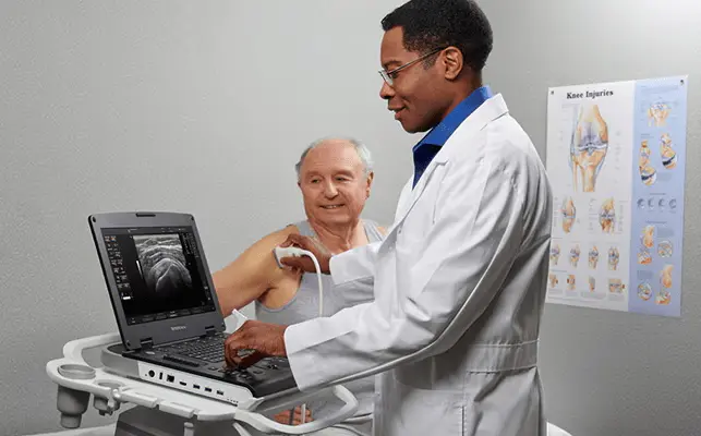
To send images to the cloud Tricefy wirelessly to share with other doctors for consultation or to share them with patients. ** / ***
** Requires Wifi connectivity and high-speed may not be available in all countries.
*** Solution for the exchange of cases based on the cloud provided separately by Trice Imaging. Customers can try out Tricefy through the trial period to sign an agreement with Trice Imaging. Trice Imaging is the sole responsibility of the application Tricefy Uplink and the cloud solution Tricefy.
AutoBladder (Vejiga Whiz)
Combines optimized imaging with automated bladder measurements for faster assessments.
Scan Assistant
Predefined exam protocols guide technologists through each step of the imaging process, reducing keystrokes, stress, and exam variability.
QTVI
Leverage raw data from TruScan to perform in-depth analysis and quantification of left ventricular function.
Images of speed tissue (TVI)
Estimated and coded by colours speeds in the tissues.
Versana Active Technology InSite™
Advanced technology of remote service of GE and diagnostics, and repair of remote monitoring; connect instantly to your ultrasound machine with a services expert GE to resolve many issues remotely in the act.
Effective follow-up examination
For follow-up examinations, the tracking tool automatically identifies the same configuration of the ultrasound probe, preset, depth, gain, and frequency that was used in the previous scan. The ultrasound image of current in real time is shown next to the image above in order to facilitate the comparison.
Thyroid Productivity
To evaluate the thyroid during the scan; to describe the characteristics of the structure in the comments that are directly incorporated in the report.
ScanCoach
This reference tool provides animated images in 3D, anatomical illustrations and images of reference of ultrasound to help determine the level of exploration suitable for the acquisition.
Needle Recognition
Perform biopsies accurate with technology to clarify the precise location of the tip of the needle.
Doppler myocardial
Images with overlay of colors in the image of the tissue.
SonoBiometry
Suggests caliper placements for standardized fetal measurements (BPD, HC, AC, FL, LH), enhancing obstetric productivity.
Breast Productivity
Provides systematic breast exam protocols, integrates BI-RADS comments directly into reports, and uses Auto Contour for easy measurement.
EF automatic
Perform an automatic tracking of the deformation of the myocardial tissue and calculate the ejection fraction of the left ventricle. The system makes the paths of the views apical four-chamber and apical two-chamber and calculates the ejection fraction.
Color B-Flow and B-Flow
B-Flow is intended to provide a more intuitive of the hemodynamic not quantitative in the vascular structures.